Saturday, April 23, 2011

Avascular Necrosis of the Knee

Avascular Necrosis

Avascular Necrosis is a disease that develops from the temporary or permanent loss of the blood provided to the bones. Bone tissue needs blood otherwise it will die causing the bone to breakdown. This can occur from trauma, non-traumatic or even pressure within the bone. Other names for Avascular necrosis include ischemic bone necrosis, osteonecrosis and aseptic necrosis.
There are also other sites for avascular necrosis to occur such as the femur, shoulder, upper arm bone and ankles. The disease may impact just one bone, multiple bones at the same time or multiple bones at separate times
Normally after an injury, a bone can heal itself by breaking down and restoring itself along with the old bone that gets drawn in and is exchanged with new bone, however if this doesn't take place and the bone is losing blood circulation then Avascular Necrosis presents itself.
At the beginning of this disease, a patient may not have any indication that they have Avascular Necrosis but as it advances the patients encounter joint pain. The pain first starts in the joint when weight is placed on the joint and then it continues on even in a relaxed state. The pain begins slowly and steadily increases to severe. The severity of this pain can restrict a persons range of motion.
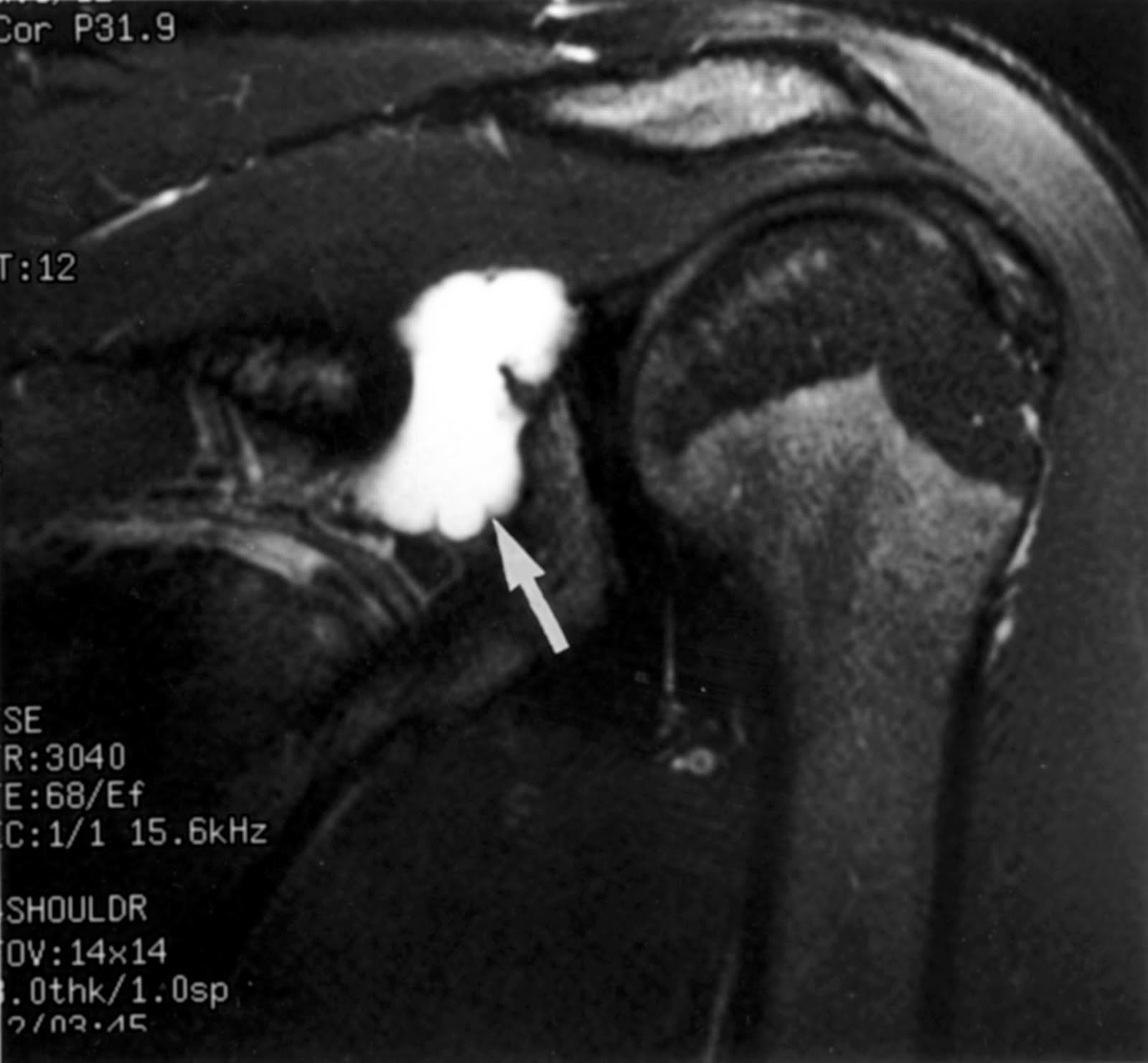
After an orthopaedic doctor has completing a physical exam and has assessed the patients history, they can determined what tests are needed to diagnose the patient as having Avascular Necrosis. X-rays can help decide if more test need to be performed to evaluate if further test are required for their diagnosis. Other diagnostic tests include MRI, CT, Bone Scan, Biopsy and Functional Evaluation of Bone.
To choose the most suitable treatment, the doctor takes into consideration the patients age, at what stage the disease is in, where the affected bone is located and its size, and what is the primary cause of Avascular Necrosis. Conservative treatment such as corticosteroid is usually given to patients first, however, these hardly ever give permanent improvement. So, more than likely the patient will have to have surgery.

Sunday, April 10, 2011

Boxer's fracture


Boxer's Fracture
A boxer's fracture is break in the bones in the knuckle region of the hand. The knuckle is the head of the metacarpal bone and the break is just below it at the neck of the metacarpal bone. The most frequent metacarpal bone to fracture when striking a fixed object is the small finger known as the fifth metacarpal. This fracture can also occur in the fourth metacarpal but some doctors include the second and third metacarpal in the explanation of a boxer's fracture.
Common symptoms of a boxer's fracture are tenderness or pain in a particular location in one of the metacarpal bones on the hand near the knuckle. The hand may appear deformed, bruise or enlarge throughout the location that is injured. If the fracture has a cut, it may be a more serious break and should be checked out as soon as possible.
Any hand injury that has symptoms or signs demonstrating a fracture should be assessed by a doctor. If you are unable to see a doctor right away, you should go to the emergency department at a hospital. The doctor will deem whether an x-ray is necessary after an examination. Before you are able to see a doctor, there are steps you can do to take care if the injury. To reduce swelling and pain, apply a cold pack or something cold to the area that is injured and elevate the broken hand to minimize the swelling. If the injured hand is cut, you run the risk of infection so it is important to wash the hand with soapy water and then conceal it promptly with a sterile bandage to decrease the chance of infection. Keep the injured hand restrained to keep from damaging nearby blood vessels, ligaments, tendons, muscles and nerves.
Once the injured hand is diagnosed with a boxer's fracture, the doctor will determine how to treat the hand and how to take care of it once you go home. If the fracture does not require surgery, a cast or splint is used to keep the hand stable. Usually with any broken bone, a person will have pain so it is important to receive some form of pain management. Pain relievers and anti-inflammatory medicine provide great alleviation. It is important to make sure that the broken bone is rehabilitating properly so it is best to look into acquiring an orthopedic specialist.


Saturday, March 19, 2011

Ganglion Cyst of the Shoulder


Ganglion Cyst
A Ganglion Cyst is a mass or lump that develops on or beneath the skin. Ganglion cyst are not only seen at the shoulder joint but are also found around the joints of the elbow, knee, hip, ankle, and foot and even more frequently, these cyst are detected on the wrist and fingers. Tissues encompassing certain joints distend with a jelly-like substance.
Recurrent injuries or moderate sprains can aggravate and break up the thin membrane that coats the tendon that causes the jelly-like substance to drain into a sac that inflates and forms a ganglion. Ganglion cyst tend to impede range of motion which hinders the joints ability to bend or straighten causing aching, irritation or soreness to develop.
An examination is typically performs through physical examination along with either an ultrasound, x-ray or MRI. If necessary, fluid may be removed from the cyst to be examined.
Treatment for some ganglion cysts is not required, however, some cysts call for Acetaminophen for minimal pain and others cysts call for Steroids for more intolerable pain. For the more bothersome cysts, surgery can be performed to remove the cyst.

Sunday, February 27, 2011

Congenital Hip Dysplasia

Congenital Hip Dysplasia

Congenital hip dysplasia is a hip joint deformity that exists at birth. The femoral head is not secure in the acetabulum and the ligaments at the hip joint may be detached or stretched. This malformation may well be hereditary but some children will present little or no traits and must be determined by a physical examination of the hip joint. Visible signs in children include walking with a limp, walking on toes and waddling stride. Characteristics consist of uneven leg positions, inconsistent fat folds, hip dislocation and decreased movement on the affected hip.

There are a few methods to diagnose congenital hip dysplasia. X-ray films help discern abnormalities of the hip joint allowing correct positioning of the hip joint if a cast is needed. Ultrasound is even more helpful because it permits multiple positioning of the hip joint. The Barlow method is where the doctor places the infant's hips together with the knees totally bent then the doctor places their middle finger over the outside of the hipbone as the thumb is positioned on the medial side of the knee. The hip is pulled away from the body where the doctor can feel whether the hip is sliding in and out of the joint. The Ortolani test is where the doctors hands surround the infants knees, with the second and third fingers turn down toward the infants thigh. Moving the legs apart, the doctor will be able to notice a pronounced clicking noise with the movement.

Exercise programs can be used to strengthen hips and help with hip motion and relieve pain along with medication as nonsurgical treatments. Treatment is needed to return the head of the femur into the hip socket. Some infants are placed in a stiff shell cast to keep the hip in position and in older children surgery may be performed to reposition the hip and then a cast is applied to keep the hip in place. Later on in life, a total hip replacement surgery may be necessary.

Sunday, February 13, 2011

Hiatal Hernia

Hiatal Hernia

A hiatal hernia transpires when a portion of the stomach pushes upward through the diaphragm. The diaphragm has a small orifice (hiatus) that permits the esophagus to move through to join the stomach. The stomach can force up through the orifice and create a hiatal hernia.
Most of the time a little hiatal hernia will not even be noticed until you go to see a doctor and he discovers it while you are being checked for something else. Taking care of yourself or taking medication such as acid reducing medicine can typically reduce symptoms. A huge hiatal hernia can cause acid and food to back flow into the esophagus, bringing on belching, heartburn, nausea and chest pain. A large hiatal hernia occasionally requires surgery to pull the stomach down into the abdomen and making the orifice in the diaphragm smaller, rebuilding a weak esophageal sphincter, or eliminating the hernia sac .

A hiatal hernia could be caused by being born with an atypically large hiatus, a genetic weakness in the nearby muscles, injury to the region, and the continuous and acute pressure on the encompassing muscles from vomiting, coughing, or during lifting hefty objects. Hiatal hernia is more typical in people who smoke, who are obese or who are age 50 or older.

Saturday, January 29, 2011

Aneurysm

Aneurysm
Aneurysm is where a section of an artery or cardiac chamber has an atypical widening or bulging attributed to weakness in the wall.

What causes aneurysms is unclear. Some aneurysms are congenital and some aneurysms come from defects in pieces of the artery wall or from trauma. Cholesterol buildup in the arteries may cause aneurysm formations and high blood pressure can increase your risk certain forms of aneurysms. Most aneurysms take place in the aorta. If it arises in the chest it is called a thoracic aortic aneurysm and if it occurs in the abdomen it is called an abdominal aortic aneurysm.

Aneurysms usually produce no symptoms but if they burst, high heart rate, lightheadedness and low blood pressure may arise resulting in a high probability of death from dangerous bleeding inside the body. Early diagnosis and medical care can help fend off many cases of ruptures and dissections. If found early, aortic aneurysms can often be treated effectively with surgery or medicines that lower blood pressure and ease blood vessels. Aortic aneurysms that are large can be braced or restored with surgery.

Atelectasis

Atelectasis
Atelectasis is brought on by an obstruction of the air passages or by compression on the outside of the lung which causes part or all of the lung to collapse. The collapse of the lung tissue impedes the respiratory interchange of carbon dioxide and oxygen.

Some risk factors for developing atelectasis are extended bed rest with small changes in position, shallow breathing, anesthesia, mucus that closes off the airway, lung diseases, foreign object in the airway, an increase of fluid between the ribs and the lungs and tumors that block the airway may lead to atelectasis. Some symptoms of acute atelectasis include cyanosis, dyspnea, elevation of temperature, shock, or a dip in blood pressure and other symptoms include c
hest pain, cough and difficulty breathing.

To diagnose atelectasis, a clinical exam is performed, a bronchoscopy to view the airways and chest x-ray. To treat atelectasis, the collapsed lung tissue needs to re-inflate. If there is fluid that is constricting the lungs then the fluid will need to be expelled so the lungs can inflate. Some treatments include postural drainage, percussion on the chest to break up mucus, deep breathing exercises, and use inhaled medications to clear the airway.

The collapsed lung typically re-inflates slowly once the blockage has been removed. A small area of atelectasis in the lung is not life threatening since the rest of the lung can compensate for the collapsed area but a large area may be life threatening, particularly in an infant or small child, or for those who have an illness or another lung disease.

To prevent atelectasis, continue deep breathes after anesthesia, urge movement and deep breathing when bedridden for an extended time, and remove small objects from the reach of small children.


Sunday, January 9, 2011

Introducing myself - RADT 415CT/MRI Procedures II

Hello all,
My name is Rhoda Stanton and I am on my third semester of clinicals for my MRI certification. I live in Indianapolis Indiana and I have worked at Methodist Sports Medicine/The Orthopedic Specialist also in Indianapolis Indiana for 10 years this coming May. I am planning on doing my clinicals again at CDI at the south office in Greenwood Indiana.
I am ready to get more quality time in at CDI and I hope to keep learning as much as possible from the great technologist there. I saw some very interesting cases at CDI last semester and I am looking forward to seeing more this semester. I hope everyone has a great semester and I am excited to reading your blogs!
Good Luck to all of you!